Doctor: Philip Gutin
Specialty: Neurosurgery
Location: Memorial Sloan-Kettering
Dr. Gutin: This tumor looked like a mushroom, with the stem inside the bone in the ear canal and the cap extended out toward the brain. They’re called acoustic neuromas and are actually benign, but they can cause neurological problems by putting pressure on the brain. The patient, a 41-year-old banker and father of two boys, was diagnosed in April 2010, after he started to experience right-side hearing loss. An ENT doctor discovered a 2.5-centimeter tumor growing inside his head.
We always tell patients that our job is to take as much of the tumor out as possible. If we have to, we’ll leave a little of the tumor behind if it’s attached to the facial nerve. You don’t want to fight with the facial nerve. The morning of surgery, the patient was brought into the OR and anesthetized. It’s a long procedure. Over five hours. It’s such a complex surgery we do it as a partner surgery, with one brain surgeon and one ear surgeon.
The first incision went behind the ear. Then we drilled a hole in the base of the skull and enlarged it.Once we removed a portion of his skull, we looked at the structure of the brain through the microscope to see the vessels and nerves. The tumor itself looked sort of yellow and soft, and we gradually teased it off, using a tool that pulverized and vacuumed it away. It’s the damnedest thing. Four hours can go by, and you have no idea.
We saw that the tumor was fairly stuck to the facial nerve, so we knew we’d have to leave a small portion behind. When most of it had been removed, we closed everything back up. The patient is recovering, day by day. It’s an incredible privilege that he trusted me enough to do this. Nobody deserves this kind of trust. It’s insane the amount of courage it takes to have your brain operated on.
As told to Katie Charles.
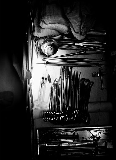
A surgical instrument tray. The tools laid out horizontally in the center of the tray are brain dissectors; the thick-handled instrument up by the sponges is for scraping bone. Photo: Pari Dukovic
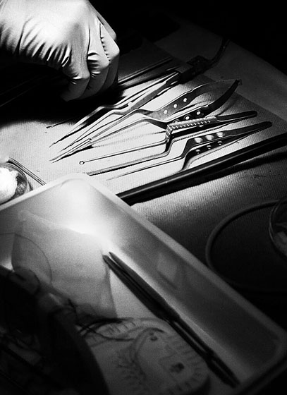
Specialized instruments for use under the microscope. Photo: Pari Dukovic
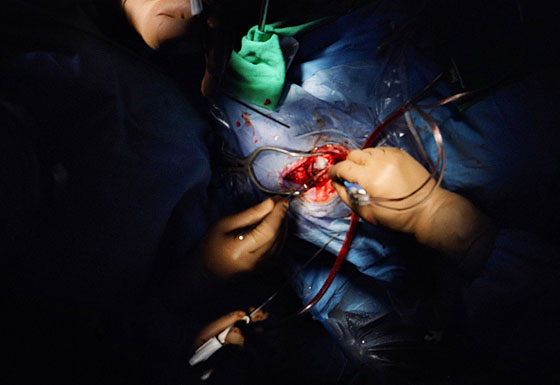
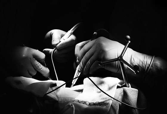
After the incision has been made, a retractor (which looks like a large tuning fork) holds the scalp and muscle back while a bipolar coagulation device (the wired device in the rightmost hand) is used to close off blood vessels. Photo: Pari Dukovic
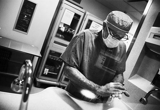
Dr. Gutin lathering his hands before entering the operating room, where the brain has already been uncovered. Photo: Pari Dukovic
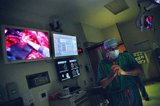
Dr. Gutin, mid-surgery, gazing at a computer guiding system, which shows the contours of the patient’s brain. Photo: Pari Dukovic

The image on the screen shows a brain retractor (big, flat, and black) pulling back the cerebellum, as well as an ultrasonic aspirator (with a whitish tip, which vibrates to scramble the tumor before suctioning it up). “It’s like a tumor vacuum cleaner,” Dr. Gutin says. Photo: Pari Dukovic
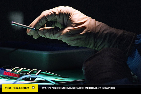
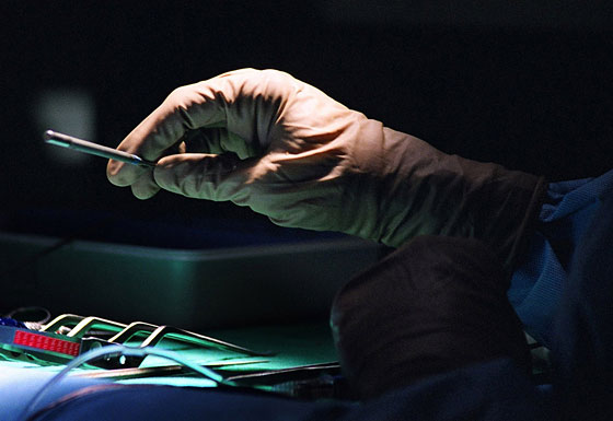
The surgeon, wielding an instrument. Photo: Pari Dukovic

Dr. Gutin assisting ear surgeon Dr. Samuel Selesnick, a partner in the operation, who is seated at the microscope. Photo: Pari Dukovic
A scrub nurse, passing an instrument. Photo: Pari Dukovic
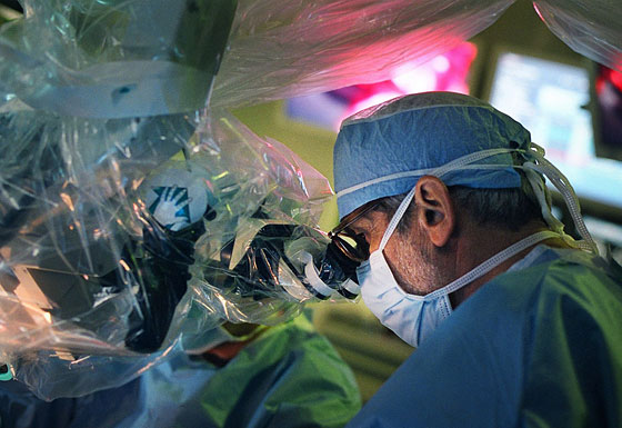
Dr. Gutin sitting at the robotic-armed microscope, with Dr. Selesnick assisting. Photo: Pari Dukovic
Dr. Gutin at the microscope. Photo: Pari Dukovic
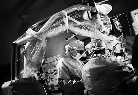
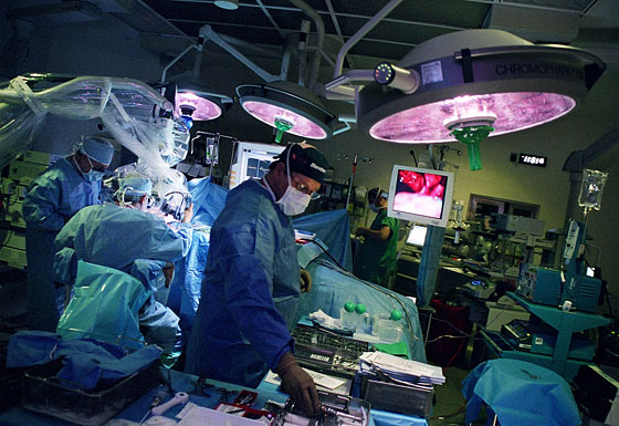
The surgeons at work. The windmill-like device on the right is actually a navigation system of sorts, and allows the doctors to know exactly where they are on the MRI scan, which was taken the day before. “It’s almost like a GPS device,” Dr. Gutin says. Photo: Pari Dukovic
The patient’s wife embraces Dr. Gutin after the surgery. Photo: Pari Dukovic