Photographs © Stanley B. Burns, M.D., and The Burns Archive
It’s been said that until 1920 or so, you were probably just as well off staying at home, letting your immune system do its work, as you were going to a hospital. Especially in the era before germ theory, when many surgeons didn’t so much as wash their instruments between patients, medical intervention could be as dangerous as disease. Yet even with their crude tools and vague knowledge, doctors and nurses did offer a measure of help and sometimes were able to do more good than harm.
Starting on August 8, Steven Soderbergh’s Cinemax series The Knick will attempt to put viewers on the scene in a New York hospital circa 1900, horse-drawn ambulances and all. Toward that goal, the show has retained as consultant Dr. Stanley Burns, keeper of the world’s greatest collection of early medical photography. The images on these pages are the tiniest sampling of the Burns Archive, which comprises more than a million photographs packed into his Murray Hill brownstone. The 40-plus books Dr. Burns has published, on themes from Victorian funeral portraits to early oncology, are sometimes horrifying, more often mesmerizing, and always a reminder that in medicine, as in no other field, nostalgia is anything but gauzy.
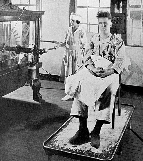
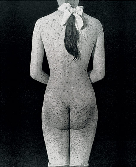
Ca. 1900
Electrotherapy. Small (and sometimes not so small) jolts were used against everything from blindness to constipation.
Photo: ” Stanley B. Burns, M.D., and The Burns Archive
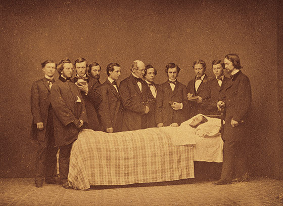
Ca. 1858
The earliest known photo of New York hospital doctors at bedside.
Photo: ” Stanley B. Burns, M.D., and The Burns Archive
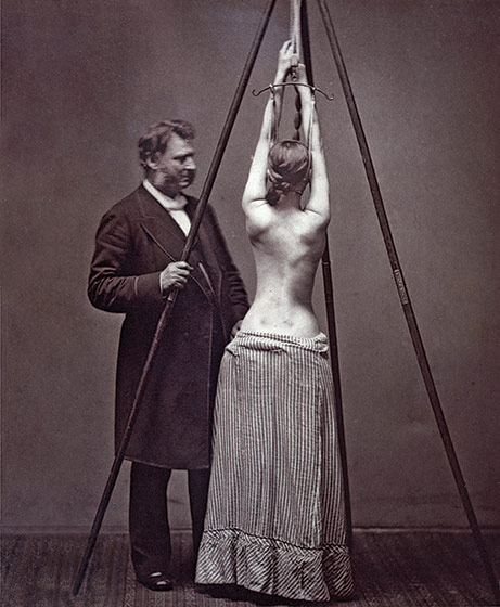
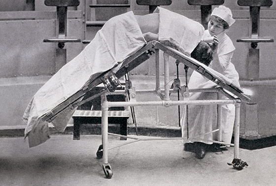
1877
Scoliosis treatment at Bellevue by
Dr. Lewis Sayre. (Once stretched, the young woman was encased in a rigid plaster cast.)
Photo: ” Stanley B. Burns, M.D., and The Burns Archive
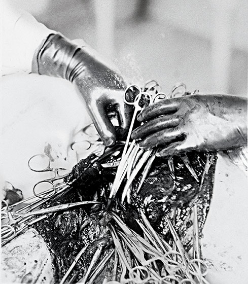
1911
Mid-mastectomy, from a step-by-step photographic guide for surgeons-in-training, produced by Johns Hopkins Hospital.
Photo: ” Stanley B. Burns, M.D., and The Burns Archive
1910
Flexing a patient’s torso in preparation for kidney surgery. The idea was to improve access to the organ by popping it out through the incision.
Photo: ” Stanley B. Burns, M.D., and The Burns Archive
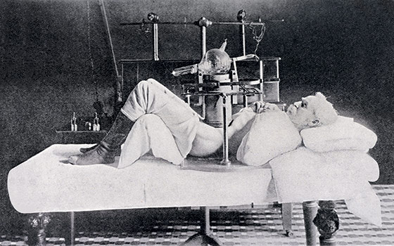
Ca. 1902
This man’s abdomen is compressed to focus
the weak output of an early X-ray tube
(that’s the glass globe at center).
Photo: ” Stanley B. Burns, M.D., and The Burns Archive